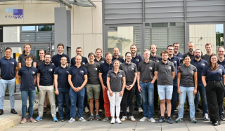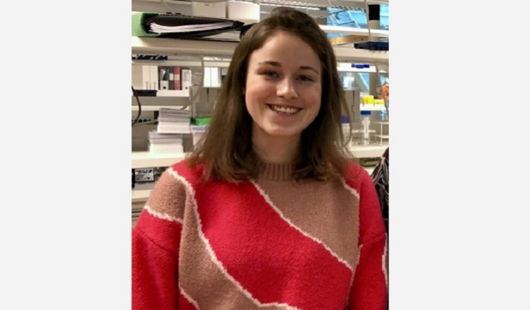
Manon, Renaud, Lucy. These three doctoral students from the Wallonia-Brussels Federation met in Uppsala, Sweden, on the sidelines of a scientific mission organised by the WBI Research and Innovation Department. Meeting with Manon Dausort.
Manon Dausort could have been a veterinarian, but she decided to study engineering sciences. Now a doctoral student at UCLouvain, last February she took part in the extensive equine mission in Scandinavia organised by WBI's Research and Innovation Department.
A mission centred on horses is a bit surprising for a civil engineer specialising in biomedical engineering, is it not? "Not really," she says. "I have been riding for over 15 years. But what brought me here concerns medical imaging and artificial intelligence."
"During my studies, I worked on the micro-changes that occurred in the brains of alcoholics when they observed short periods of abstinence. A non-invasive medical imaging technology was used to detect these changes."
"When patients entered the hospital, we would have them undergo a structural ("diffusion") MRI scan. They were given a second examination on discharge a few days later. This was to demonstrate a possible recovery of their brain function, their white matter, as well as the connectivity in the brain."
From the brain to malignant tumours
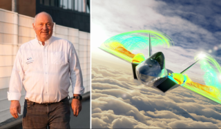
Since last year, as part of her PhD at the PiLAB (Pixels and Interactions) of Prof. Benoît Macq (UCLouvain), and under the aegis of the Trail Institute, she has been using the same tools, but this time applying them to tumours. This is to better characterise cancer cells and follow their evolution over time.
"Right now, I am focusing on a review of the literature on the subject. I also analyse the functioning of the screening tools used to determine their weaknesses and limitations. At the same time, in the laboratory, we are trying to improve the performance of the diffusion MRI used for white matter imaging, with a view to making it equally effective for examinations of other organs. Especially for breast cancer screening."
Why focus on breast cancer? "We know that mammography screening delivers good results," she says. "A biopsy is performed if there is any suspicion. This kind of invasive examination is difficult for patients, especially psychologically. By improving medical imaging, for example by using diffusion MRI, our goal is to better detect and characterise possible suspicious tumours so we can limit the need for a biopsy. We want to develop a complementary tool, which will be used by doctors."
Earlier detection
"Doctors base their diagnosis on a visual examination of mammograms. They also have a classification tool. At a certain threshold, they decide to perform a biopsy. The tool we want to develop is a more neutral and impartial model. The idea is to improve prevention tools, including very early in the development of a tumour, as soon as a few malignant cells appear, before they form a certain mass."
"In theory, what we want to develop could be applied to any part of the body," continues Manon Dausort. "We chose the breast because the preventive mammography examination is based on an X-ray technique. Our tool, diffusion MRI imaging, is based on the use of a magnetic field, not ionising radiation."
Her doctoral project is based on medical imaging technology, as well as on data analysis using artificial intelligence. So what is the link with horses, which were the focus of the exchanges organised in Sweden? "Simply because the technologies I am working on could also find applications in the veterinary field, and particularly in the equine field," she says, by way of conclusion.
This article was written by Christian Du Brulle for the Daily Science platform, with the support of Wallonia-Brussels International.