Christian Du Brulle, from the Daily Science platform, met Renaud Van Damme as part of a mission organized by the Research & Innovation Department of Wallonia-Brussels International in Sweden. Check out his interview.
In the auditorium of SLU, the Swedish University dedicated to agricultural sciences based in Uppsala, Renaud Van Damme is all smiles. "I learned that a Belgian scientific delegation (organised by the Research and Innovation Department of Wallonia-Brussels International) was coming to visit the veterinary faculty. So I didn't hesitate for a moment. It was a great opportunity to meet and talk to Walloon researchers."
The Belgian doctoral student, originally from Jumet, began his higher education at the technical campus of the Haute École in Hainaut (Mons), where he completed a Bachelor's degree in biotechnology with a focus on bioinformatics. During this time, he had the opportunity to complete an Erasmus residency at SLU in 2018. He has hardly left the veterinary faculty of the Swedish university since.
Bovine metagenomics
"The prospect of being able to take my studies further at Uppsala first took the form of a Master's degree," he explains. "I then had the opportunity to secure funding for a PhD." As a result, he has been living in Uppsala for five years now. And does he speak Swedish? "Not really," he confesses. "Everything is done in English here."
For Renaud Van Damme, the most important issue is elsewhere. It is bioinformatics that holds his attention. "As part of my PhD, I analyse and develop tools and protocols related to metagenomics," he explains.
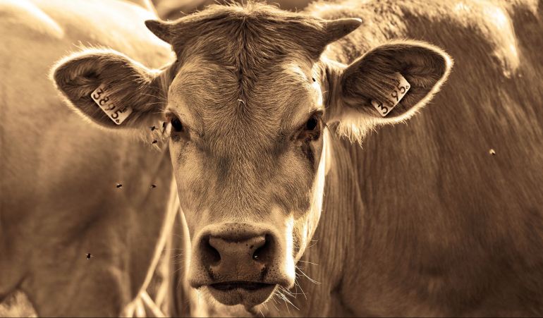
"I study the microorganisms present in different environments and I try to determine the importance of their presence as well as their interactions and functions. This is mainly in bovine stomachs. The idea is to better understand the interactions between the bacterial populations present in these digestive systems and the cow breeds."
The composition of microbiota as a subject of research
The bioinformatician's work focuses on cattle breeds raised in Ethiopia and South Africa. "We are trying to understand the interactions that may exist between the animal's genome and the species of microorganisms linked to it," he says.
"More specifically, I am working on the correlations between variations in the genome of these animals and the composition of their microbiota. We know that the composition of microbiota depends on many factors, such as the mother's microbiota, the environment and the food. Personally, I focus on the genetics of the animal. Because even if we act on a whole series of external parameters, such as a change in the diet or the use of antibiotics, we notice that the composition of the animal's microbiota returns to its initial composition after a while."
The researcher has already developed several analysis tools during the first years of his doctoral studies. Now he needs to use them on his samples. "This has been delayed due to Covid-19, as well as the civil war that is ravaging Ethiopia. My samples only arrived at the lab recently," he says. But he is confident. He knows his techniques and already has laboratory practice.
Previously, the researcher was able to familiarise himself with molecular genetic analysis techniques on a few rare white (and even albino) elk from Sweden. The idea was to try to determine which mutant genes could be responsible for this situation.
Coffee breaks and cinnamon pastries
When you look out of the windows of the veterinary faculty in Uppsala, the snow that covers a large part of the landscape reminds visitors that we are much further north than Charleroi, Mons or Jumet. But this does not put Renaud Van Damme off. On the contrary. The quality of life, the contact with students and the efficiency of public transport have won over the doctoral student. When the young man talks about his five years in Sweden, he immediately mentions the appeal of "fikas".
These are the traditional coffee breaks with cinnamon pastries. Coffee breaks, of course, but also moments that have become almost institutionalised in the country and encourage social gatherings.
"During fikas, it's easy to talk to colleagues from different departments that we don't necessarily meet during our work, or to have informal but often very interesting discussions with someone," says Renaud Van Damme. He then concludes, "At the university, it's clear that fikas encourage good science."
Find all of Christian Du Brulle's articles on the Daily Science platform, with the support of Wallonia-Brussels International.